The micro-XRD/XFM beamline will provide:
- Simultaneous X-ray fluorescence (XRF) and X-ray diffraction (XRD) rapid mapping measurements not conceivable on laboratory-based equipment;
- Monochromatic and Laue diffraction (white or pink light);
- Phase mapping, strain mapping, crystallite orientation (all 2D or 3D via DAXM) and composition (2D);
- Selected area X-ray absorption spectroscopy (XAS, both XAFS and XANES) for determination of local coordination and/or oxidation state;
- Spot size adjustable from below 1 µm and up to 5 µm.
When the crystallite size is considerably smaller than the beam size monochromatic diffraction measurements yield either complete or fragmented Debye-Scherrer diffraction rings. These rings offer considerable information, readily enabling phase identification, with preferred orientation derivable from intensity variations and the ring width being a function of strain and crystallite size. However, monochromatic radiation has the disadvantage that where the crystallite size is larger than the beam (as is becoming more often the case due to on-going improvements in focussing optics to considerably less than 1 μm) few or no diffraction peaks may be measurable for a given sample and detector geometry. Thus to offer a truly versatile microdiffraction capability which is applicable to all crystallite sizes white or pink light diffraction must also be available.
Diffraction patterns, generated from high intensity broad band pass synchrotron radiation, may be produced from deep within a sample. Deconvolution of diffraction patterns as a function of depth can be carried out by differential aperture X-ray microscopy (DAXM). The 3D information that can be derived from Laue DAXM data includes crystallite sizes, crystallite orientation, definition of grain boundaries and crystallite deviatoric strain i.e. distortion of the unit cell. The ability to measure these characteristics at micron scale is central to understanding and developing material’s properties. It is the 3D structural capability, coupled to XRF and XAS that will ensure that the micro-XRD/XFM will be a world leading facility.
The Australian scientific community does not currently have ready access to imaging microdiffraction synchrotron facilities and thus the provision of the micro-XRD/XFM will truly be a new and important resource. The potential impact of synchrotron microdiffraction coupled with XRF and XAS on the fundamental and applied sciences has been recognised across a wide cross-section of the Australian scientific community comprising earth scientists, biologists, metallurgists, forensic scientists and materials scientists. Official recognition of the need for a microdiffraction facility has also been provided in the Synchrotron Strategic Plan 2007-2017 where this was named as being one of the four facilities that should be implemented at the Australian Synchrotron in the near future.
A survey of the potential utilisation by only a limited number of institutions comprising the University of South Australia, University of Adelaide, Curtin University of Technology, Southern Cross University, La Trobe University, CSIRO (Minerals Division, Ensis Joint Venture, Energy Storage Group, Land and Water Division, Exploration and Mining Division, Molecular Health and Technologies Division, Materials Science and Engineering Division) AMIRA International Ltd. (representing over 60 national and international mining, mineral processing, smelting and chemical/equipment supply companies) and ANSTO indicates immediate over subscription from these institutions alone.
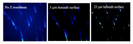
From left to right, Laue (white light) X-ray diffraction images of nickel metal with no depth resolution and depth resolved at 5 μm and 25 μm. The ‘streaks’ in the first two images indicate the presence of strain in the near surface region, while the well defined ‘spots’ in the third image indicates a much lower level of strain in the bulk material (supplied by Dr. Gene Ice and recorded at the 34ID-E beamline, APS).
Refs: i B.C. Larson, W. Yang, G.E. Ice, J.D. Budai, J.Z. Tischler, Three-dimensional X-ray structural microscopy with submicrometre resolution, Nature, 415, 887-890 (2002).
ii G.E. Ice, B.C. Larson, J.Z. Tischler, W. Liu, W. Yang, X-ray microbeam measurements of subgrain stress distributions in polycrystalline materials, Materials Science and Engineering A, 399, 43–48 (2005).
 Download MMC Detailed Report 2011-09 (pdf, 622kb)
The members of the MMC Beamline Scoping Group are: Andrea Gerson (UniSA), Steve Wilkins (CSIRO), Brian Abbey (Melbourne), Peter Lynch (Deakin) and Qinfen Gu (AS).
Enquiries may be directed to: Andrea Gerson
Position: Director ACeSSS, University of South Australia
Phone: +61 (0)8 8302 3044
Email: Andrea.Gerson@unisa.edu.au
Further Information
- Contact Andrew Peele or Kia Wallwork at asdp@synchrotron.org.au.