 Accurate knowledge about the distribution of transition metals such as calcium, iron, manganese and zinc could help resolve key medical, biological and environmental questions such as how much atmospheric carbon dioxide could be sequestered within the deep waters of the earth’s oceans.
Martin de Jonge from the Australian Synchrotron and his local and US collaborators recently announced a significant improvement in elemental imaging that will provide unprecedented detail of the three-dimensional distribution of elements in a wide range of sample types.
Called x-ray fluorescence tomography, the technique creates three-dimensional x-ray fluorescence ‘maps’.
X-ray fluorescence microscopy is a proven technique for obtaining valuable elemental, structural and chemical information. The Australian Synchrotron’s x-ray fluorescence microprobe (XFM) can resolve details as small as around one-thousandth the diameter of a typical human hair. It can also detect various elements at much lower concentrations than laboratory-based techniques such as proton-induced x-ray emission.
The technique is often used to produce two-dimensional maps by projection imaging – much the same as you might obtain for a broken arm. In this kind of imaging, all of the parts are cast as a shadow onto the film, essentially flattening out the patient or object. While useful, this imaging mode loses the three-dimensional nature of the object, and with it goes much valuable information. Tomography is a technique that can be used to infer the 3-D distributions from 2-D projection images; when the doctor uses this it is called C-T or a CAT scan.
Martin and co-workers have developed the techniques of three-dimensional computed tomography and applied them to the problems of fluorescence imaging. In particular, they have improved the resolution of the measurement to below 400 nanometres. Their measurements were taken at the APS in July 2008, over 32 hours of beamtime.
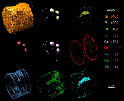
Martin and his colleagues used the new technique to produce 3-D x-ray fluorescence images of a freshwater diatom. In addition to the detail they obtained for known structures inside the diatom, the researchers made some surprising new findings about iron and manganese distribution in association with silicon at submicron length scales. These preliminary findings appear to suggest that iron and manganese may play a role in silica deposition.
In addition to these developments at the APS, Martin and the team at the XFM beamline are developing the techniques of x-ray fluorescence tomography at the Australian Synchrotron. The technique has been tested using the Kirkpatrick-Baez microprobe and the Maia detector, and has achieved an estimated 3-D resolution of around two micrometres. Current upgrades to the XFM scanning stages will improve scanning performance for 2-D and 3-D work, and will make this one of the world’s premier facilities for micrometre fluorescence studies.
‘Definition’ refers to the number of pixels used to represent an image. Previous implementations of fluorescence tomography have been limited to low-definition by data acquisition times. The present work increases the resolution by a factor of 10 and the definition by a factor of greater than 1000 over previous work. Martin says that such measurements will become routine in the near future with present trends in data acquisition and detector sensitivity.
The collaborators reported their work in the Proceedings of the National Academy of Sciences: Martin D. de Jonge, Christian Holzner, Stephen B. Baines, Benjamin S. Twining, Konstantin Ignatyev, Julia Diaz, Daryl L. Howard, Daniel Legnini, Antonino Miceli, Ian McNulty, Chris J. Jacobsen, and Stefan Vogt, 2010, Quantitative 3D elemental microtomography of Cyclotella meneghiniana at 400-nm resolution, PNAS vol 107 no.36, 15676-15680
Image: These images are 3D renderings of elemental distributions in a freshwater diatom (Cyclotella meneghiniana).
Isosurface concentrations used for the display are indicated in mmol/L. Clear correspondences between P (phosphorus), K (potassium), and Ca (calcium) are observed in the organelles, with a small amount of Mn (manganese) apparent. The Si (silicon) frustules and the Fe (iron) and Mn (manganese) rings are striking; and the cytoplasmic pillar (running along the axis of the diatom) contains mainly Cl (chlorine), Cu (copper), Zn (zinc), and S (sulphur - not shown). As with the concentration thresholds shown here, total elemental content spans three orders of magnitude, ranging from 8 μg for Si (silicon) to 2 ng for Mn (manganese). From PNAS 107, 15676-15680, with permission.