A technique that creates ‘virtual’ sections of small 3D samples is now available for general user proposals on the x-ray fluorescence microscopy beamline.
Thanks to intensive local development work with the Maia detector on the XFM beamline, fluorescence tomography is no longer limited to measuring very small objects at low resolution. Maia’s speed and sensitivity enable users to obtain tomographic images of elemental distribution within a specimen without the long exposure times and high x-ray doses previously associated with fluorescence tomography. For delicate samples, this substantially reduces the risk of x-ray damage occurring before measurements can be obtained, as illustrated by a recent study of metal distributions in fresh fully-hydrated plant roots [1,2].
General users can apply to use fluorescence tomography from round 2012/1 (January-May 2012). A strong science case will be an important part of your proposal, and users are urged to discuss their requirements with beamline staff prior to submission.
 The sinogram to the left shows measurements obtained from a fully hydrated, immature grain of rice. Three sinograms are superimposed: the white, determined from the Compton scatter signal, maps the grain’s biological ultrastructure. The red sinogram shows germanium, which also serves as a proxy for Si, and the green sinogram maps zinc.
The reconstructed 2D section of the rice grain is shown below (scale bar = 600 μm). The reconstruction resolution is around 5 micrometres, with the husk clearly visible around the outside of the grain. Despite localised germanium and zinc hot-spots, their distribution is still clearly elucidated. Germanium inhabits the hairs on the outside of the husk and some structures within the grain. Zinc is found in both the endosperm and the husk.
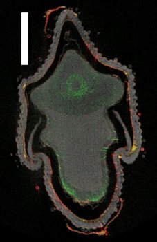 In rice, arsenite (the dominant arsenic species in paddy fields) is taken up and transported through the silicic acid pathway. Enzo Lombi (Centre for Environmental Risk Assessment and Remediation, University of South Australia) and his colleagues investigated germanium in this specimen as a way of indirectly investigating the distribution of silicon. Germanium can be analysed with hard x-rays and is amenable to tomography in large samples; while silicon’s low fluorescence energy means it cannot be efficiently investigated with this technique due to self-sorption.
In the edible part of the grain, germanium was found mainly in the ovular vascular trace, which is the structure that delivers nutrient to the grain. This distribution closely follows arsenite distribution in the grain, suggesting that silicic acid transporters are not only responsible for the plant taking up arsenite but also for uploading this contaminant in the rice grain. Zinc was found in the embryo and in the aleurone layer.
During the three hours required for this measurement, the specimen would not have been subjected to any higher x-ray dose than that experienced during the equivalent 2D measurement from an actual cross-section.
Although the maximum size of the specimen that can be measured is no longer limited by hardware capabilities, self-absorption – the absorption of the x-ray fluorescence as it leaves the specimen – prevents measurement of arbitrarily-sized specimens. As a guide, absorption of the fluorescence should be less than about 50% over the specimen thickness in order for a measurement to be feasible. Refer to [3] for a detailed discussion, or contact Martin de Jonge martin.dejonge@synchrotron.org.au at the XFM beamline for advice.
[1] E Lombi, MD de Jonge, E Donner, et al.: Fast X-Ray Fluorescence Microtomography of Hydrated Biological Samples. Plos One 6 (2011)
[2] PM Kopittke, NW Menzies, MD de Jonge, et al.: In Situ Distribution and Speciation of Toxic Copper, Nickel, and Zinc in Hydrated Roots of Cowpea. Plant Physiology 156 663 (2011)
[3] MD de Jonge & S Vogt: Hard X-ray fluorescence tomography - an emerging tool for structural visualization. Current Opinion in Structural Biology 20 606 (2010).