Selenium is a dietary puzzle. If there isn’t enough selenium in your diet, you’re more likely to suffer from some forms of cancer. On the other hand, if there’s too much selenium in your diet, you may suffer a range of adverse effects. The different chemical forms of selenium in the diet appear to play a big role in its impact on our bodies.
In October 2011, an international team led by Adelaide researchers revealed important new details of how selenium begins to fight cancer cells.
Hugh Harris and his collaborators used x-ray techniques at the Australian Synchrotron, and at overseas facilities through the synchrotron’s international access program, to reveal what happens to selenium when it enters human lung cancer cells in inorganic selenite form.
Hugh and his colleagues found that the selenite was quickly taken up by cancer cells but then took a day or more to start killing them.
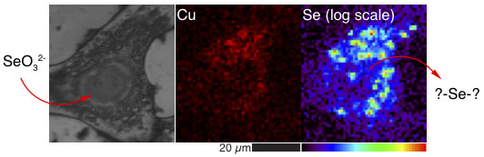 Left: XRF elemental maps of human cancer cells treated with selenite show that selenium and copper are stored together in small compartments. Images: Hugh Harris, University of Adelaide.
During the first 24-48 hours, the inorganic selenite is transformed through several intermediate forms into a different chemical form of selenium that indicates oxidative stress in the cancer cells. Oxidative stress is a major area of research into a number of diseases.
The synchrotron results also showed that lung cancer cells store selenium in specific areas that contain raised copper levels as well, which may indicate a link between the effects of dietary selenium and copper.
The aim of the project is to learn how the chemical form of selenium changes over time in human lung cancer cells and use this information to assist the development of better ways to use selenium to treat human cancers while reducing the risk of adverse health effects.
Hugh and his University of Adelaide colleagues are doing this work in collaboration with researchers from the University of Sydney, Argonne National Laboratory in the US, and the Australian Synchrotron. The work is part of a broad, long-term investigation of selenium metabolism, storage, accumulation and general biology, and how selenium interacts in the body with other metals such as copper and iron.
At the Australian Synchrotron, the team used micro-x-ray absorption near edge structure (micro-XANES) and x-ray fluorescence microscopy (XFM) to identify the specific chemical forms of the selenium and where these were located within the cancer cells.
Click here for more information about the group’s October 2011 research paper in the Journal of the American Chemical Society.
Click here to read an earlier article about Hugh’s work from Lightspeed June 2010.