A promising experimental radiotherapy technique that uses finely divided synchrotron x-rays to destroy tumours without seriously affecting normal tissue is under investigation at the Australian Synchrotron.
Around half of all cancer patients receive radiotherapy. Although effective, radiotherapy treatments still have significant limitations. For example, the radiation dose delivered to a tumour can’t exceed the dose that can be tolerated by normal tissues surrounding the tumour.
The experimental technique known as microbeam radiation therapy (MRT) challenges pre-conceived ideas about radiotherapy for two main reasons. Firstly, normal tissue seems to tolerate peak MRT doses 100 times greater than the doses used in conventional radiotherapy. Secondly, entire tumours may be destroyed under MRT even though only one-tenth of their volume has been irradiated at peak doses.
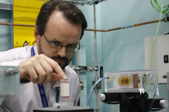
Photo above: Jeff Crosbie from The University of Melbourne places a tumour sample on the IMBL beamline for x-ray irradiation.
Microbeam radiation therapy uses a lattice of kilo-voltage x-ray ‘micro-beamlets’. The beamlets are 100-300 microns apart (100 microns is roughly the width of a human hair) and each beamlet is 10-50 microns wide. Typical in-beam radiation doses are 500-1000 Gray, and doses in the valleys between the microbeams are approximately 5-20 Gray.
Much of the ground-breaking work with MRT has been carried out at the European Synchrotron Radiation Facility (ESRF) in France. The imaging and medical beamline (IMBL) at the Australian Synchrotron has been constructed to enable radiotherapy trials in animals and humans, and recently underwent several months of further commissioning and characterisation work.
Jeff Crosbie from the University of Melbourne is part of an Australian research team using the imaging and medical beamline and the recently-completed near-beam preparation area inside the main Australian Synchrotron building. Jeff and other researchers from The University of Melbourne, led by Professor Peter Rogers, together with colleagues from CSIRO and The Alfred Hospital conducted MRT experiments on the IMBL in December 2011.
To investigate the ability of tumour cells to form viable colonies after irradiation with synchrotron microbeams, the researchers irradiated flasks of breast tumour cells with high-dose microbeam radiation therapy using the beamline’s new MRT collimator and shutter system. The team also collected data they will use to determine which genes are active in tumour cells at different times following microbeam irradiation. The group are busy analysing their data; just in time for grant-writing season.

Photo above: Monica Sharma, a 2011 honours student at The University of Melbourne, processes an irradiated tumour sample.