New images show how ‘cell death’ protein gets ready for action
A new discovery by some of Australia’s best medical researchers could lead to more effective treatments for some types of cancer and neurodegenerative diseases.
The Melbourne-based researchers have obtained highly detailed pictures of a key molecular mechanism in the process of apoptosis, also called programmed cell death. This process governs the turnover of cells in our body. If the process doesn’t work properly, the results can include the growth of cancer cells – which have essentially lost the instructions that tell them to die, or the death of too many neurons – which can lead to the development of neurodegenerative conditions.
Researchers from the Walter and Eliza Hall Institute of Medical Research in Melbourne used x-ray diffraction at the Australian Synchrotron to obtain highly detailed, three-dimensional images of the activation of a protein called Bax. The synchrotron x-ray images show how Bax is activated when small proteins fragments bind to it, and how the activated Bax proteins bind to each other to form molecular complexes that can punch holes in cell membranes.
 The work is described in a paper published in February 2013 in a major scientific journal called Cell. The research was carried out by Walter and Eliza Hall Institute’s Dr Peter Czabotar, Professor Peter Colman and Dr Dana Westphal and their colleagues.
Dr Czabotar said the synchrotron x-ray images were “really exciting”. The Bax images are the latest discovery and a major highlight in several years of work on the Australian Synchrotron’s specialised x-ray diffraction equipment.
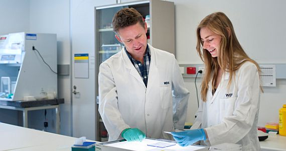
Photo at right: Peter Czabotar and Dana Westphal in the laboratory. Photo: WEHI
Click here for the Australian Synchrotron media release.
Click here for the Walter and Eliza Hall Institute media release.