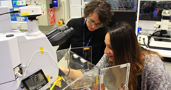
Associate Professor Natalie Sims, leader of the research team, with Christina Vrahnas, first author on the Bone journal paper.
Melbourne researchers have used the Australian Synchrotron to reveal how bone made in people taking hormone treatment for advanced osteoporosis is likely to be stronger and more durable than previously thought, offering new insights into skeletal diseases and ways of predicting who may be at risk of fractures and breaks.
The experimental technique, developed by researchers from St Vincent’s Institute of Medical Research (SVI) in Melbourne and revealed online in Bone ahead of appearing in the December edition, uses infrared synchrotron light to monitor tiny sections of newly-formed bone at different times, in conditions mimicking the human body, influenced by parathyroid hormone treatment (PTH) or teriparatide.
SVI’s Associate Professor Natalie Sims, leader of the research team, says PTH is one of very few recommended second-line treatments for people with serious bone diseases, including osteoporosis.
‘PTH stimulates the production of new bone when bone fragility persists after first-line treatments fail, but previous studies suggested this replacement bone was not as strong as existing bone which, in turn, was thought to be behind ongoing fractures and breaks in people who had daily PTH injections.
‘Not only has our analysis shown replacement bone has exactly the composition and structure that it should, our investigative technique opens the door to previously impossible bone examination, which could shed new light on all skeletal diseases including osteogenesis imperfecta and osteomalacia.’
Associate Professor Sims says using the Australian Synchrotron, landmark research infrastructure of the Australian Nuclear Science and Technology Organisation (ANSTO), gave the research team new appreciation of how bones regrow, in unprecedented detail and accuracy.
‘When bone forms, collagen is laid down in a matrix, in which calcium builds over time in a process called mineralisation – it is crucial that we can probe deep into the bone matrix because on the surface, just like wood, both weak and strong bone can look exactly the same.
‘Moreover, the ability to analyse tiny bone sections only 15 microns across – about the size of two blood cells – meant we could go deeper and deeper into older and older bone in tiny increments, to identify structural differences: clues as to which bone was truly strong and which was truly weak.’
Professor Peter Ebeling AO, Head, Department of Medicine, Monash University at Monash Health says the new avenue of bone research could lead to improved diagnostic tests and approaches to predicting breaks and fractures.
‘Current bone density tests are quite good at telling us who is at most risk of fractures but, in many cases, they do not tell the full story.
‘By looking at tiny samples, as opposed to centimetres-long bone sections, it will be tremendous if this research can help inform a more precise and useful approach, as well as assisting with testing the efficacy of future, improved bone disease drugs that are able to build new bone.’
Media coverage
- 'Useful break in bone study', Herald Sun, Friday 28 October 2016